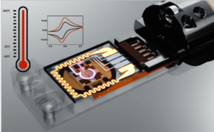
Inspiration

Bruker FlatQUAD

Where Conventional SDDs Reach Limitations.

Have you ever thought about what element mapping would be like if you could do fast mapping down to a few nanometers?

The unique annular design of the Bruker XFlash® FlatQUAD detector and its position between the SEM pole piece and the sample lead to an unmatched solid angle which directly translate into faster measurements. Together with the ESPRIT software suite, the QUANTAX FlatQUAD provides the ideal instrument to analyze e.g. sensitive samples at lowest beam currents or samples with high topography.

The XFlash® FlatQUAD can also turn your SEM or FIB into low voltage analytical STEM to analyze electron transparent samples at highest spatial and spectroscopic resolution in a more timely and cost-efficient way.

Protochips – AXON

Do you know exactly where the beam has been on your sample?
Do you know exactly how much dose has been injected in every one of these locations?
Would you like to know?

Protochips AXON and AXON Dose can do this and much more.
AXON redefines the in situ experience by linking the transmission electron microscopy detectors and in situ systems together with a revolutionary new software platform. It improves data quality, enhances and extends your current microscope capabilities and makes in situ experiments easier for the novice to most advanced users.

With accurate calibrations using semi-automated machine vision routines, AXON Dose ensures the best accuracy and precision for dose quantification.

Bruker QUANTAX ED-XS

Bruker QUANTAX ED-XS is a new product package combining the new and unique eFlash XS EBSD detector with the robust XFlash® EDS detector under the versatile ESPRIT software suite. This hardware and software combination is a new approach to providing powerful analytical tools to the larger microscopy community which has not been offered EBSD capabilities until now.

Bruker Optimus 2

Brukers new, augmented TKD solution builds on the unmatched performance of existing on-axis TKD through the addition of multiple new hardware options, accessories, and software features. Most important change is the release of OPTIMUS 2 detector head.

Fischione Model 1061

Fischione Model 1061 – Advanced sample preparation
For many of today’s advanced materials, analysis by SEM is an ideal technique for rapidly studying material structure and properties.

Fischione Instruments’ Model 1061 SEM Mill is an excellent tool for creating the sample surface characteristics needed for SEM imaging and analysis.

Bruker Hysitron In -Situ Nano-mechanics testing

As the world leader in nanomechanical testing systems, Bruker makes it easy for you to conduct in-situ mechanical experiments in your electron microscope (SEM/FIB/TEM) with the Hysitron PI Series PicoIndenters.

Our unique transducer design delivers unmatched stability throughout your experiments, resulting in precise data even at the nanoscale. Video capture from the microscope enables real-time monitoring and direct correlation of mechanical data to microscope imaging. With solutions designed to fit many of the microscope brands in use, you are sure to find one that is ideally suited to your research.

Spicer Magnetic Field Cancelling System

Electron microscopes have to operate in an ambient magnetic field comprising the earth’s field and fields radiated by electric power networks and electrical machines. When the ambient field changes, the electron beam in the microscope is deflected, causing loss of resolution and image distortion.

Spicer Consulting magnetic field cancelling systems protect sensitive equipment in the world’s leading laboratories, universities, and semiconductor manufacturing plants, as well as in the test facilities of electron and ion beam equipment manufacturers. Its magnetic field.

Protochips - In Situ TEM Solutions

The Protochips in situ systems are designed to accelerate your research by enabling dynamic, real-time visualization of nanoscale phenomena. Explore our suite of TEM solutions.

The Atmosphere system creates an in situ reaction chamber inside your TEM microscope, enabling atomic resolution imaging of dynamic nanoscale processes under realistic conditions.

Poseidon Select creates a miniature liquid cell inside the TEM. It enables you to visualize numerous nanoscale processes in their native environment such as corrosion, particle analysis, biological materials and battery materials.

Fusion Select transforms your TEM into an in-situ laboratory using the flexibility of MEMS-based E-chips that deliver accurate, precise experimental conditions.

NanoMegas-PRECESSION ELECTRON DIFFRACTION

Precession electron diffraction (PED) is a specialized method to collect electron diffraction patterns in a transmission electron microscope (TEM). By rotating (precessing) a tilted incident electron beam around the central axis of the microscope, a PED pattern is formed by integration over a collection of diffraction conditions. This produces a quasi-kinematical diffraction pattern that is more suitable as input into directmethods algorithms to determine the crystal structure of the sample.

Rulla till toppen